Digital X-Rays
Digital X-rays (radiographs) are a valuable diagnostic tool in veterinary medicine. As we continually strive to offer the highest quality medicine and diagnostic testing, we are pleased to offer digital radiographs as a means of providing excellent care to our patients.
A digital radiograph is a type of computer-generated photograph that can look inside the body and reveal information that may not be discernable from the outside or from the veterinary exam.
Digital radiographs:
Digital radiographs can evaluate almost any organ in the body and can be shown to the client within minutes. Radiographs can be used to evaluate bones as well as the size, shape, and position of many of the body's organs. The size of organs is important because some medical conditions such as kidney, heart, or liver disease, can alter the size of these organs. The shape and position of organs can be altered or distorted by certain medical conditions, including intestinal blockage or cancer. Tumors, depending on their size and location, can also sometimes be detected. Radiographs can also be used to diagnose bladder stones, broken bones, chronic arthritis, certain spinal cord diseases, and a variety of other conditions.
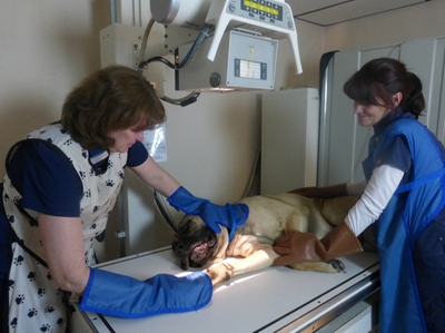
Radiographs are painless, safe, and completely non-invasive. We sometimes need to sedate patients simply because positioning can be difficult with large dogs or because the dog or cat is painful. Because the level of radiation exposure needed to perform radiographs is very low, even pregnant females and very young pets can have the procedure performed.
Radiographs are an important tool that can help us make a correct diagnosis for your pet. Our radiology service is staffed by caring and skilled professionals who will provide state-of-the-art care with compassion and expertise. Digital images are permanently stored in the patients' records and can be sent to specialists as needed very easily.
A digital radiograph is a type of computer-generated photograph that can look inside the body and reveal information that may not be discernable from the outside or from the veterinary exam.
Digital radiographs:
- Reveal high levels of detail when assessing internal structures like the heart, lungs, abdominal organs, and bones
- Use lower dosages of radiation than conventional radiographs
- Are better for the environment because no chemicals are needed to process silver-based films
- Take less time because the images are available immediately
Digital radiographs can evaluate almost any organ in the body and can be shown to the client within minutes. Radiographs can be used to evaluate bones as well as the size, shape, and position of many of the body's organs. The size of organs is important because some medical conditions such as kidney, heart, or liver disease, can alter the size of these organs. The shape and position of organs can be altered or distorted by certain medical conditions, including intestinal blockage or cancer. Tumors, depending on their size and location, can also sometimes be detected. Radiographs can also be used to diagnose bladder stones, broken bones, chronic arthritis, certain spinal cord diseases, and a variety of other conditions.
Radiographs are painless, safe, and completely non-invasive. We sometimes need to sedate patients simply because positioning can be difficult with large dogs or because the dog or cat is painful. Because the level of radiation exposure needed to perform radiographs is very low, even pregnant females and very young pets can have the procedure performed.
Radiographs are an important tool that can help us make a correct diagnosis for your pet. Our radiology service is staffed by caring and skilled professionals who will provide state-of-the-art care with compassion and expertise. Digital images are permanently stored in the patients' records and can be sent to specialists as needed very easily.